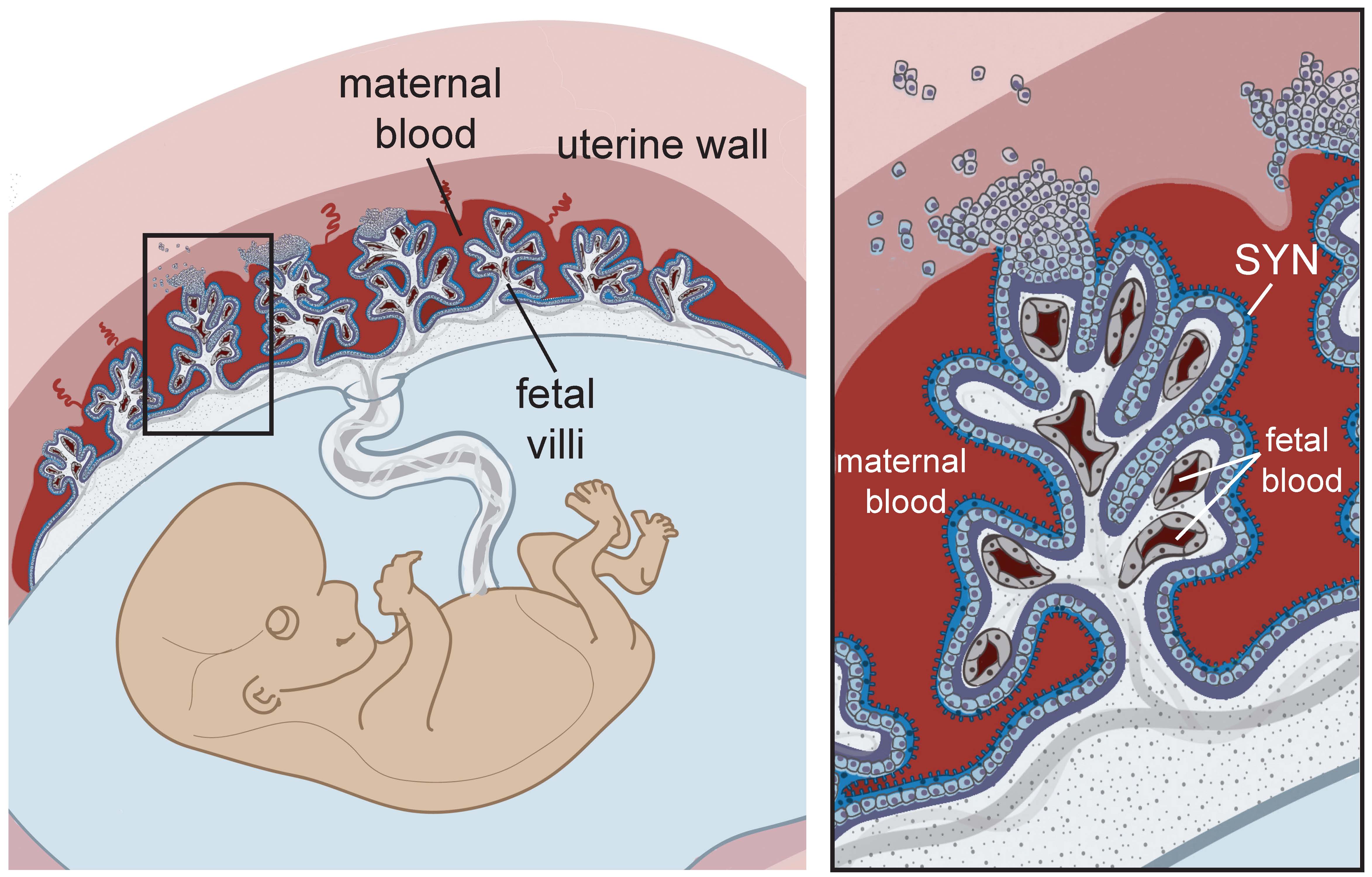
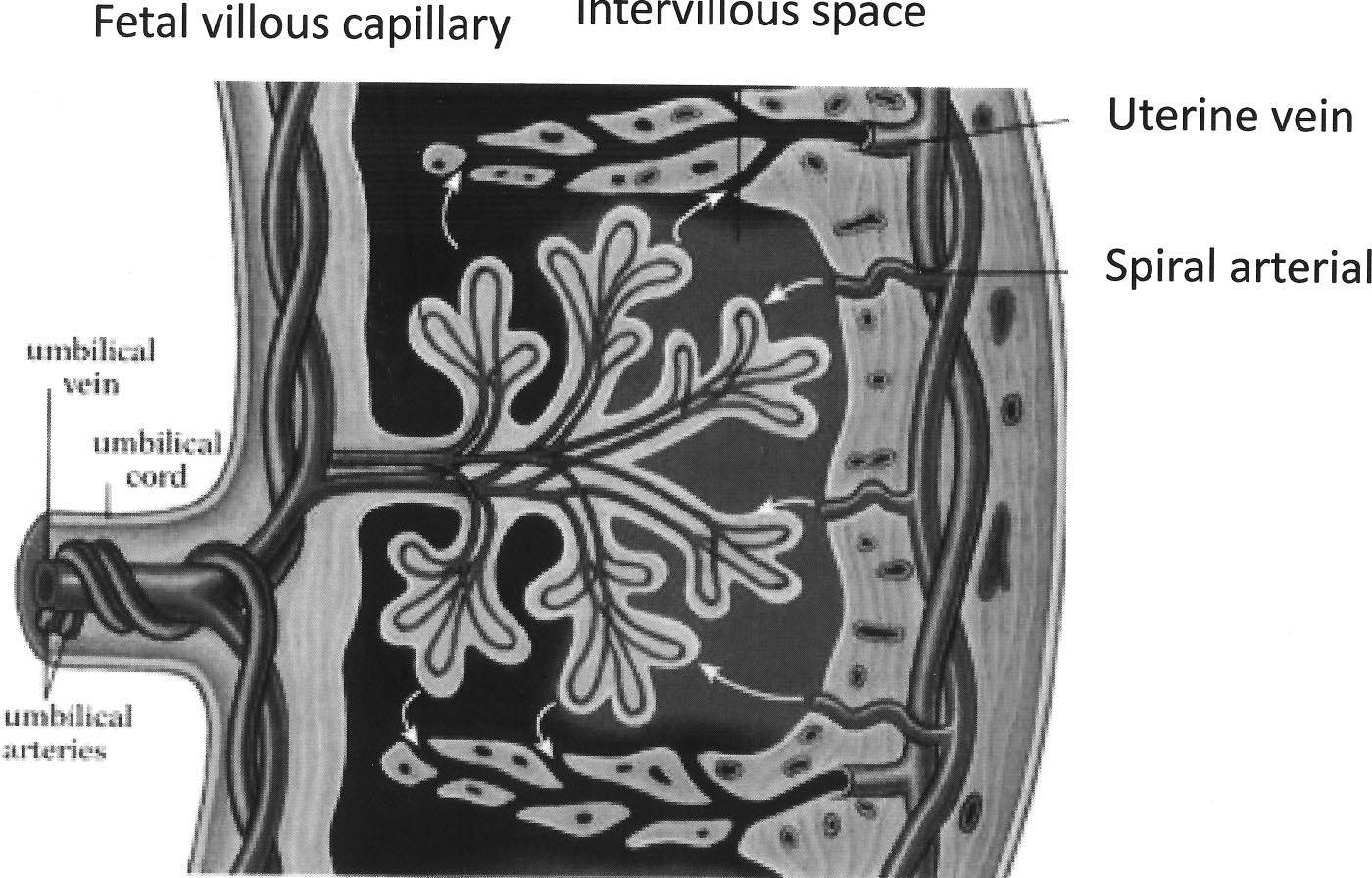
Placental structure and circulation at term Maternal blood enters Biology Diagrams The placenta is arguably the most important organ of the body, but paradoxically the most poorly understood. These are not displaced until onset of the fetal placental circulation towards the end of the first trimester. Leitner W, Anderson M, Friedman JE, Draznin B. 2004. Human placental growth hormone increases expression of the p85 The placenta is a unique vascular organ that receives blood supplies from both the maternal and the fetal systems and thus has two separate circulatory systems for blood: (1) the maternal-placental (uteroplacental) blood circulation, and (2) the fetal-placental (fetoplacental) blood circulation. The uteroplacental circulation starts with the maternal blood flow into the intervillous space The placenta is the medium through which material passes from the maternal circulation to the fetal circulation by passive diffusion or active transport.[1] The placental growth factor is released from the placenta to prepare the mother's body for pregnancy in terms of cardiovascular adaption. (HCS), also known as human placental

Most of the circulation to the lower body is supplied by blood passing through the ductus arteriosus. This blood then enters the umbilical arteries and flows into the placenta. In the placenta, carbon dioxide and waste products are released into the mother's circulatory system, and oxygen and nutrients from the mother's blood are released into
Blood Circulation in the Fetus and Newborn Biology Diagrams
During the pregnancy the uterine circulation constantly adapts in order to be adequate for the growing metabolic needs of the embryo. Via the spiral arteries (80 -100 mm Hg) that come from the uterine arteries (Aa. uterinae), maternal blood gets into the intervillous spaces in a region delimited by the anchoring villi.

Development of the placental vasculature. (A) Placental villi of 6 weeks gestational age prior to onset of the chorionic circulation, showing the presence of nucleated erythrocytes in the developing fetal capillaries (arrowed).(B) Villi at 14 weeks gestational age showing the presence of non-nucleated erythrocytes in the larger vessels within the stromal core, indicative of onset of the
Placenta Circulation - an overview Biology Diagrams
The feto-placental circulation continues to develop structurally and mature functionally throughout gestation as a result of continuing growth and maturation of villous trophoblast 9, whereas the structural remodeling of the utero-placental vasculature is considered to be complete shortly after 20-22 weeks of human pregnancy.
